Monitoring and Documentation
|
AUTHOR: Jeff Gadsden Introduction The incidence of complications from general anesthesia has diminished substantially in recent decades, largely due to advances in respiratory monitoring. (1) The use of objective monitors such as pulse oximetry and capnography allows anesthesiologists to quickly identify changing physiologic parameters and intervene rapidly and appropriately. In contrast, the practice of regional anesthesia has traditionally suffered from a lack of similar objective monitors that aid the practitioner in preventing injury. Practitioners of peripheral nerve blocks were made to rely on subjective end points to gauge the potential risk to the patient. This is changing, however, with the introduction and adoption of standardized methods by which to safely perform peripheral nerve blocks with the minimal possible risk to the patient. For example, instead of relying on feeling "clicks," "pops," and "scratches" to identify needle tip position, the anesthesiologist can now directly observe it using ultrasonography. It follows that advancements such as this may help in reducing the three most feared complications of peripheral nerve blockade: nerve injury, local anesthetic toxicity, and inadvertent damage to adjacent structures ("needle misadventure"). Objective monitoring, and the rationale for its use, is discussed in the first part of this chapter. The later section focuses on documentation of nerve block procedures, which is a natural accompaniment to the use of these empirical monitors. The proper documentation of how a nerve block was performed has obvious medicolegal implications and aids the future practitioner in choosing the best nerve block regimen for that particular patient. SECTION I: MONITORING What are the Available Monitors? 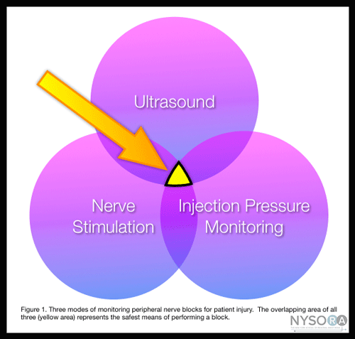 Figure 1: Three modes of monitoring peripheral nerve blocks for patient injury. The overlapping area of all three (yellow area) represents the safest means of performing a block. Monitors as used in the medical sense, are devices that assess a specific physiologic state and warn the clinician of impending harm. The monitors discussed in this chapter include nerve stimulation, ultrasonography, and the monitoring of injection pressure. Each of these has its own distinct set of both advantages and limitations. For this reason, these three technologies are best used in a complementary fashion (Figure 1), to minimize the potential for patient injury, rather than just relying on the information provided by one monitor alone. The combination of all three monitors is likely to produce the safest possible environment in which to perform a peripheral nerve block. A fourth monitor that many clinicians use regularly is the use of epinephrine in the local anesthetic. Good evidence supports this practice as a means of improving safety during peripheral nerve blocks, particularly in patients receiving higher doses of local anesthetic. First, it acts as a marker of intravascular absorption. About 10 to 15 μg of epinephrine injected intravenously reliably increases the systolic blood pressure >15 mm Hg, even in sedated or beta-blocked individuals (whereas a heart rate increase is not reliable in sedated patients). (2,3) The recognition of this increase permits the clinician to halt the injection promptly and increase his or her vigilance for signs of systemic toxicity. Second, epinephrine truncates the peak plasma level of local anesthetic, resulting in a lower risk for systemic toxicity. (4,5) Concerns regarding the effects of epinephrine on nutritive vessel vasoconstriction and nerve ischemia have been unsubstantiated. In contrast, concentrations of 2.5 μg/mL have been associated with an increase in nerve blood flow, likely due to the predominance of the beta effect of the drug. (6) Therefore, when added to local anesthetics, epinephrine can enhance safety during administration of larger doses of local anesthetics. Nerve Stimulation Neurostimulation has largely replaced paresthesia as the primary means of nerve localization in the 1980s and has only recently been challenged by ultrasound guidance. Its effectiveness as a method of nerve localization has been challenged since the publication of a series of studies showing that, despite intimate needle-nerve contact as witnessed by ultrasonography, a motor response may be absent. (7) In some instances, a current intensity as high as >1.5 mA may be necessary to elicit motor response with needle placement within epineurium of the nerve. (8) There are probably multiple factors that contribute to the explanation of this phenomenon, including the nonuniform distribution of motor and sensory fibers in the compound nerve and the unpredictable pattern of current dispersion in the tissue depending on tissue conductances and impedances. Although this has led some clinicians to de-value nerve stimulation in an era of ultrasound-guided blocks, a growing body of evidence suggests that the presence of a motor response at a very low current (i.e., Table 1: Studies of Intensity of the Current (mA) and Needle Tip Location
More recently, a study was conducted on 55 patients scheduled for upper limb surgery who received ultrasound guided supraclavicular brachial plexus blocks. The authors set out to determine the minimum current threshold for motor response both inside and outside the first trunk encountered. (11) They discovered that the median minimum stimulation threshold was 0.60 mA outside the nerve and 0.3 mA inside the nerve. Interestingly, stimulation currents of ≤0.2 mA were not observed outside the nerve, whereas 36% of patients experienced a twitch at currents Taken together, these data suggest that although the sensitivity of a "low-current" twitch for intraneural placement is not high, the specificity is. Put another way, the needle tip can be in the nerve and not elicit a motor response at very low currents; however, if a twitch is elicited at Most regional anesthesiologists agree that injection of local anesthetic into the nerve may be a risk factor for injury and that extra-neural deposition minimizes the potential for an intrafascicular injection. (12) Ultrasonography is good, but not perfect, at delineating the exact position of the needle tip. In our attempts to get "close, but not too close" to the nerve so we might have the best block result, needles occasionally but inevitably cross the epineurium into the substance of the nerve. This event in and of itself may be of minimal consequence. (13) However, injection into a fascicle carries a high risk of injury. (14) It is for this reason that a reliable electrical monitor of needle tip position is a useful safety instrument. If a motor twitch is elicited at currents Overall, nerve stimulation adds little to the cost of a nerve block procedure, in terms of time, clinician effort, or dollars. It also serves as a useful functional confirmation of the anatomic image shown on the ultrasound screen (e.g. "Is that the median or ulnar nerve?"). In our practice, nerve stimulator is routinely used in conjunction with ultrasound guidance as an invaluable monitor of the needle tip position with respect to the nerve, based on the association of low currents with intraneural placement. In addition, an unexpected motor response during ultrasound-guided blocks may alert the operator of the needle-nerve relationship that was missed on ultrasound. Ultrasonography The use of ultrasound guidance to assist in nerve block placement has become very popular, for a number of reasons. First, ultrasound allows visualization of the needle in real time and therefore quickly and accurately guide the needle toward the target. Multiple injection techniques that were difficult, or indeed dangerous, to do in the era of nerve stimulation alone are now easy to perform because the nerves can be seen and injectate carefully deposited at various points around them. Also, because a motor response is not technically required, blocks can now be performed in amputees who do not have a limb to twitch. Not surprisingly, ultrasound has the potential to improve the safety of peripheral nerve blocks for a number of reasons. First, adjacent structures of importance can be seen and avoided. The resurgence in popularity of the supraclavicular block is a testament to this. Before ultrasound, the highly effective block was relatively unpopular as a means of anesthetizing the brachial plexus, for fear of causing a pneumothorax, despite the paucity of data regarding its actual incidence. However, now that the brachial plexus and, more importantly, the rib, pleura, and subclavian artery can all be seen at the supraclavicular level, this block has become common in clinical practice. However, recent reports of pneumothoraces serve as a reminder that while ultrasound may reduce the incidence of complications of nerve blocks, it is unlikely to entirely prevent when used as a sole monitor. (15,16) Similarly, there are reports of intravascular and intraneural needle placement witnessed (and despite the use of) ultrasound, highlighting the need to use care with this technology that is, in the end, a tool that is not failsafe. (17-19) A useful adjunct to the visualization of structures on the ultrasound screen is the ability to measure the distance from skin to target using electronic calipers (Figure 2). This, coupled with needles that have depth markings etched on the side of the nerve block needles, confers a great safety advantage by warning the clinician of a "stop distance," or a depth beyond which he or she should stop, reassess the needle visualization, and perhaps withdraw and begin again. Another important advantage that ultrasound can confer is the ability to see the local anesthetic distribution on the screen image (Figure 3). If corresponding tissue expansion is not seen when injection begins, then the needle tip is not where it is thought to be, and the clinician should immediately halt injection and relocate the tip of the needle. This is particularly worrisome in vascular areas because the lack of spread can signal the intravascular needle placement. However, ultrasound has been used successfully to diagnose an intra-arterial needle tip placement when an echogenic "blush" was noted in the lumen of the artery, allowing for rapid cessation of the block technique and avoidance of what surely would have been systemic toxicity. (20,21) 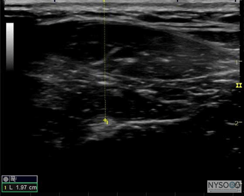 Figure 2: An example of ultrasound being used to determine the depth of a structure of interest. Similarly, ultrasound may also be able to reduce the likelihood of systemic toxicity by allowing clinicians to use less local anesthetic. Several authors have published large reductions in the volume required to affect an equivalent block to standard nerve stimulation techniques. For example, Casati et al demonstrated a significant reduction in volume required to produce an effective three-in-one block (22 mL vs. 41 mL). (22) Sandhu et al showed in a feasibility study that infraclavicular block was possible using ultrasound with volumes typically half of what were used with nerve stimulation alone (16.1 ± 1.9 mL). (23) Riazi et al published a study in 2008 showing that ultrasound guidance allowed for a substantial reduction of volume for interscalene block used for postoperative pain while still providing a quality block (5 mL vs. 20 mL). (24) Interestingly, this low dose also resulted in less diaphragmatic impairment related to phrenic nerve paresis. The utility of ultrasound in prevention of nerve injury during peripheral nerve blockade is likely over-estimated. The problem is threefold: First observing the needle tip in relation to the nerve is user dependent, and one can often be fooled by poor technique or simply unfavorable echogenic characteristics of the tissue-needle interface; second, the current resolution available is not adequate to distinguish between an intraversus extrafascicular needle tip location. This difference is crucial because evidence is mounting that an intraneural (but extrafascicular) injection is likely not associated with injury, whereas injection inside the fascicles themselves produces clinical and histologic damage. (14,25) Lastly, once injection has begun, even a minuscule amount of local anesthetic can produce damage if intrafascicular. (26) Relying on the visual confirmation of tissue expansion may result in damage before expansion is detected on the screen. It is, in other words, probably too late.
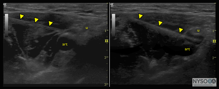
Figure 3: Axillary block with axillary artery (art), ulnar nerve (u), needle (arrowheads), (A) before and (B) after injection of small amount of local anesthetic, showing spread of injectate between artery and nerve. Injection Pressure Monitoring How, then, can the clinician distinguish the intrafascicular versus the extrafascicular needle tip placement, if ultrasound guidance is insufficient? An additional modality to ultrasound and nerve stimulation is monitoring of injection pressures. In a study of intraneural injections in dog sciatic nerves, a slow injection of lidocaine while the needle tip was intrafascicular was associated with an immediate and substantial rise in the pressure of the syringe-tubing-needle system (>20 psi), followed by return of the pressure tracing to normal (i.e., 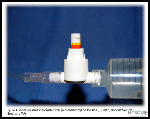 Figure 4: Inline pressure manometer with graded markings on the side (B-smart, Concert Medical, Needham, MA). The use of "hand feel" to avoid high injection pressure is unfortunately not reliable. Studies of experienced practitioners blinded to the injection pressure and asked to perform a mock injection using standard equipment reveals wide variations in applied pressure, some grossly exceeding the established thresholds for safety. (27) Similarly, anesthesiologists perform poorly when asked to distinguish between intraneural injection and injection into other tissues such as muscle or tendon in an animal model. (28) It is therefore important to use an objective and quantifiable method of gauging injection pressure. Although the practice of injection pressure monitoring during peripheral nerve blocks is relatively young, monitoring options do exist. Tsui et al described a method of "compressed air injection technique" where 10 mL of air was drawn into the syringe along with the local anesthetic. (29) Holding the syringe upright, it is then possible to avoid exceeding a maximum threshold of 1 atmosphere (or approximately 15 psi) by only allowing the gas portion of the syringe contents to compress to half of its original volume, or 5 mL. This makes use of Boyle's law, which states that pressure times volume must be constant. A pressure Another option is disposable pressure manometers specifically manufactured for this purpose. These devices bridge the syringe and needle tubing, and via a spring-loaded piston, allow the clinician to gauge the pressure in the system continuously. On the shaft of the piston are markings delineating three different pressure thresholds:20 psi (Figure 4). An advantage of this method is the ease with which an untrained assistant who is performing the injection can read and communicate the pressures. In addition, the syringe does not have to be held upright, as in the compressed air technique. Pressure monitoring may be a useful safety monitor for other aspects of peripheral nerve blocks. In a study of patients receiving lumbar plexus blocks randomized to low (20 psi) pressures, Gadsden et al demonstrated that 60% of patients in the high-pressure group experienced a bilateral epidural block. (30) Furthermore, 50% in the same group reported an epidural block in the thoracic distribution. No patient in the low-pressure group experienced bilateral or epidural blockade. This has now become an important adjunct to lumbar plexus blockade in our institution, to avoid this potentially dangerous complication. 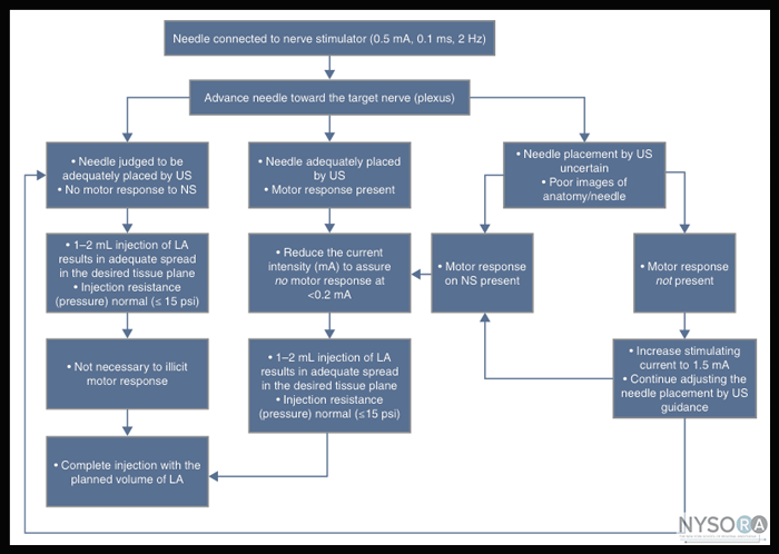
Figure 5: Flowchart depicting the order of correctly documenting nerve block procedures: combining ultrasound (US), nerve stimulation (NS), and injection pressure monitoring. Summary Regional anesthesia has been making a transition from art to science as more rigorous and precise means of locating nerves are developed. The same process should be expected for monitoring peripheral blockade. The use of neurostimulation, ultrasonography, and injection pressure monitoring together provides a complementary package of objective data that can guide clinicians to perform the safest blocks possible. The flowchart in Figure 5 outlines how these monitors can be combined. SECTION II: DOCUMENTATION Block Procedure Notes
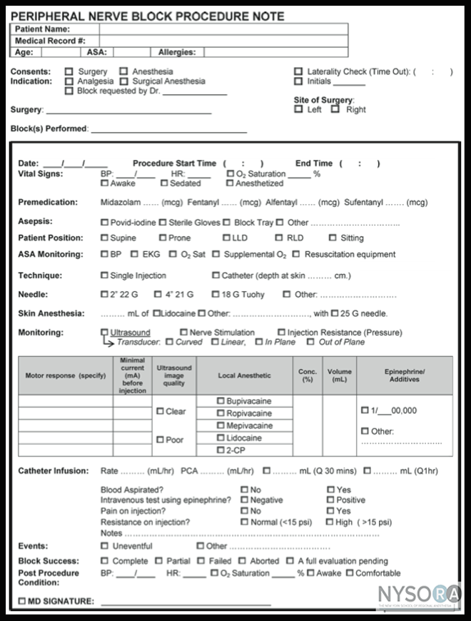
Figure 6: Example of an anesthetist procedure note. Documentation of nerve block procedures has, by and large, lagged behind the documentation of general anesthesia, and it is often relegated to a few scribbled lines in the corner of the anesthetic record. Increasing pressure from legal, billing, and regulatory sources has provoked an effort to improve the documentation for peripheral nerve blocks. A sample of a peripheral nerve block documentation form that incorporates all of the monitoring elements discussed in this book (ultrasound, nerve stimulation, and injection pressure monitoring) and can be adopted to individual practice is shown in Figure 6. This form has a number of features that should be considered by institutions attempting to formulate their own procedure note. These include the following:
Another useful aspect of peripheral nerve block documentation is the recording of an ultrasound image or video clip, to be stored either as a hard copy in the patient's chart or as an electronic copy in a database. This is not only good practice from a medicolegal point of view but is a required step that must be taken if the clinician wishes to bill for the use of ultrasound guidance. Any hard copies should have a patient identification sticker attached, the date recorded, and any pertinent findings highlighted with a marker, such as local anesthetic spreading around nerve circumferentially (Figure 6). Informed Consent Documentation of informed consent is another issue that is of importance to regional anesthesiologists. Practice patterns vary widely on this issue, and specific written consent for nerve block procedures is often not obtained. However, the written documentation of this process can be important for a number of reasons:
The following tips can be used to maximize the consent process: A specific regional anesthesia consent form should be included as well. This will not be applicable to all institutions but can be modified to suit the needs of each individual department.
|
| 02/20/2016(+ 2016 Dates) | |
| 01/27/2016 | |
| 03/17/2016 | |
| 04/20/2016 | |
| 09/23/2016 | |
| 10/01/2024 |
![[advertisement] gehealthcare](../files/banners/banner1_250x600/GEtouch(250X600).gif)

![[advertisement] concertmedical](../files/bk-nysora-ad.jpg)




Post your comment