Wrist Block
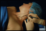
 A 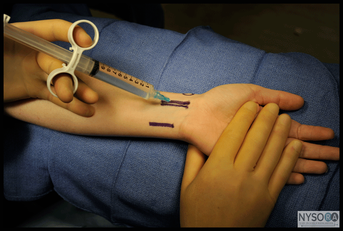 B Figure 1: (A) Technique to accomplish a wrist block. (B) Median nerve block. Needle is inserted medial or lateral to the flexor palmaris longus tendon and carefully advanced to avoid paresthesia. Then 5 mL of local anesthetic is injected. Essentials
General Considerations
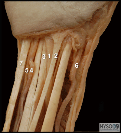
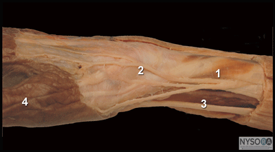
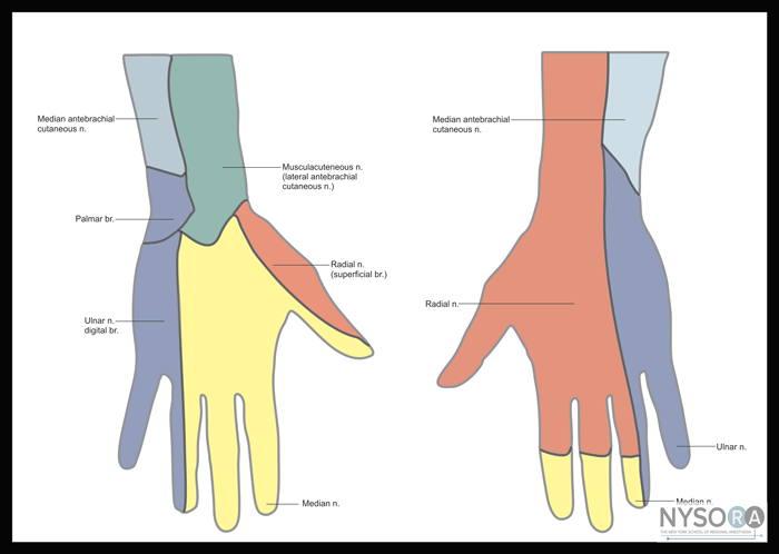
Figure 2: (A) Anatomy of the right wrist. (1) median nerve. (2) flexor palmaris longus. (3) flexor carpi radialis. (4) ulnar artery. (5) Ulnar nerve. (6) radial artery (7) flexor carpi ulnaris. (B) Anatomy of the right superficial radial nerve. (1) superficial radial nerve. (2) radial styloid. (3) flexor carpi radialis tendon. (4) thumb. A wrist block consists of anesthetizing the terminal branches of the ulnar, median, and radial nerves at the level of the wrist. It is an infiltration technique that is simple to perform, essentially devoid of systemic complications, and highly effective for a variety of procedures on the hand and fingers. The relative simplicity, low risk of complications, and high efficacy of the procedure mandates this block to be a standard part of the armamentarium of an anesthesiologist. Several different techniques of wrist blockade and their modifications are in clinical use; in this chapter, however, we describe the one most commonly used at our institution. Wrist blocks are used often for carpal tunnel and hand and finger surgery. Functional Anatomy Innervation of the hand is shared by the ulnar, median, and radial nerves (Figure 2 and 3). The ulnar nerve provides sensory innervation to the skin of the fifth digit and the medial half of the fourth digit, and to the corresponding area of the palm. The same area is covered on the corresponding dorsal side of the hand. Motor branches innervate the three hypothenar muscles, the medial two lumbrical muscles, the palmaris brevis muscle, all the interossei, and the adductor pollicis muscle. The median nerve traverses the carpal tunnel and terminates as digital and recurrent branches. The digital branches supply the skin of the lateral three and a half digits and the corresponding area of the palm. Motor branches supply the two lateral lumbricals and the three thenar muscles (recurrent median branch). Although there is significant variability in the innervation of the ring and middle fingers, the skin on the anterior surface of the thumb is always supplied by the median nerve and that of the 5th finger by the ulnar nerve. The palmar digital B branches of the median and ulnar nerves also innervate the nail beds of the respective digits. The radial nerve lies on the anterior aspect of the radial side of the forearm. About 7 cm above the wrist, the nerve deviates from the artery and emerges from the deep fascia, dividing into medial and lateral branches to supply sensation to the dorsum of the thumb and the dorsum of the hand (the first three and one-half digits as far as the distal interphalangeal joint). Distribution of Blockade Blocking the ulnar, median, and radial nerves results in anesthesia of the entire hand. The nerve contribution to innervation of the hand varies considerably; Figure 3 shows the most common arrangement. Equipment A standard regional anesthesia tray is prepared with the following equipment:
Figure 3: Cutaneous innervation of the left hand. Landmarks and Patient Positioning The patient is positioned supine, with the arm in abduction. The wrist is best kept in slight extension. 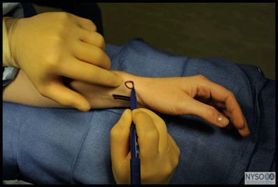
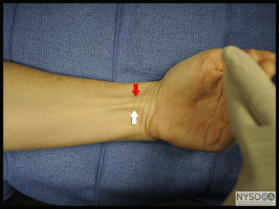
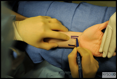
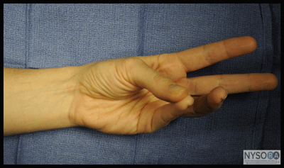
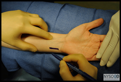
Figure 4: Palpation of the radial styloid. The superficial radial nerve is blocked by an injection just proximal to the styloid. Maneuvers to Facilitate Landmark Identification The superficial branch of the radial nerve emerges from between the tendon of the brachioradialis and the radius just proximal to the easily palpable styloid process of the radius (circle) (Figure 4). Then it divides into the medial and lateral branches, which continue subcutaneously on the dorsum of the thumb and hand. Several of the branches pass superficially over the anatomic "snuffbox." The median nerve is located between the tendons of the flexor palmaris longus (white arrow) and the flexor carpi radialis (red arrow) (Figure 5A and B). The flexor palmaris longus tendon is usually the more prominent of the two, and it can be accentuated by asking the patient to oppose the thumb and 5th finger while flexing the wrist (Figure 6); the median nerve passes just lateral to it. The ulnar nerve passes between the ulnar artery and tendon of the fl exor carpi ulnaris (Figure 7) . The tendon of flexor carpi ulnaris is superficial to the ulnar nerve.
Figure 5: A maneuver to accentuate the tendons of the flexors of the wrist. (A) Shown are flexor palmaris longus (white arrow) and flexor carpi radialis (red arrow) tendons. (B) Outlining flexor palmaris longus tendon.
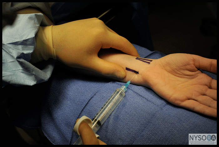
Technique The entire surface of the wrist and palm should be disinfected. Block of the Ulnar Nerve The ulnar nerve is anesthetized by inserting the needle under the tendon of the flexor carpi ulnaris muscle close to its distal attachment just above the styloid process of the ulna. The needle is advanced 5 to 10 mm to just past the tendon of the flexor carpi ulnaris (Figure 9A). After negative aspiration, 3 to 5 mL of local anesthetic solution is injected. A subcutaneous injection of 2 to 3 mL of local anesthesia just above the tendon of the flexor carpi ulnaris is advisable for blocking the cutaneous branches of the ulnar nerve, which often extend to the hypothenar area.
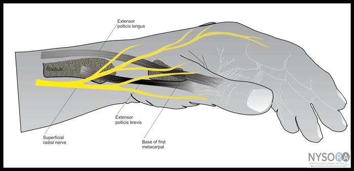
Figure 8: Common arrangement of superficial branches of the radial nerve Block of the Median Nerve The median nerve is blocked by inserting the needle between the tendons of the flexor palmaris longus and flexor carpi radialis (Figure 9B). The needle is inserted until it pierces the deep fascia, and 3 to 5 mL of local anesthetic is injected. Although piercing of the deep fascia has been described to result in a fascial "click," it is more reliable to simply insert the needle until it contacts the bone. The needle is withdrawn 2 to 3 mm, and the local anesthetic is injected.
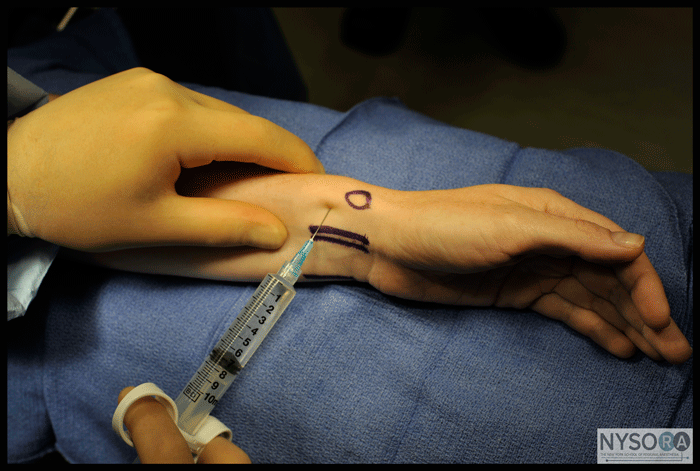
Block of the Radial Nerve The radial nerve block is essentially a "field block" and requires more extensive infiltration because of its less predictable anatomic location and division into multiple smaller cutaneous branches. Five milliliters of local anes- thetic should be injected subcutaneously just proximal to the radial styloid, aiming medially. Then the infiltration is extended laterally, using an additional 5 mL of local anesthetic (Figure 9C).
Block Dynamics and Perioperative Management The wrist block technique is associated with moderate patient discomfort because multiple insertions and subcutaneous injections are required. Appropriate sedation and analgesia (midazolam 2-4 mg and alfentanil 250-500 µg) are useful to ensure the patient's comfort. A typical onset time for a wrist block is 10 to 15 minutes, depending on the concentration and volume of local anesthetic used. Sensory anesthesia of the skin develops faster than the motor block. Placement of an Esmarch bandage or a tourniquet at the level of the wrist is well tolerated and does not require additional blockade. Complications and How to Avoid Them Complications following a wrist block are typically limited to residual paresthesias due to inadvertent intraneural injection. Systemic toxicity is rare because of the distal location of the blockade and the relatively small volumes of local anesthetics (Table 1). Table 1: Complications of Wrist Block and Techniques to Avoid Them 
|
| 02/20/2016(+ 2016 Dates) | |
| 01/27/2016 | |
| 03/17/2016 | |
| 04/20/2016 | |
| 09/23/2016 | |
| 10/01/2024 |
![[advertisement] gehealthcare](../../../files/banners/banner1_250x600/GEtouch(250X600).gif)

![[advertisement] concertmedical](../../../files/bk-nysora-ad.jpg)






Post your comment