Ultrasound Physics
|
AUTHOR: Daquan Xu Introduction Ultrasound application allows for noninvasive visualization of tissue structures. Real-time ultrasound images are inte- grated images resulting from reflection of organ surfaces and scattering within heterogeneous tissues. Ultrasound scanning is an interactive procedure involving the operator, patient, and ultrasound instruments. Although the physics behind ultrasound generation, propagation, detection, and transformation into practical information is rather complex, its clinical application is much simpler. Understanding the essential ultrasound physics presented in this chapter should be useful for comprehending the principles behind ultrasound-guided peripheral nerve blockade. History of Ultrasound 
Figure 1: Early application of Doppler ultrasound by LaGrange to perform supraclavicular brachial block. In 1880, French physicists Pierre Curie, and his elder brother Jacques Curie, discovered the piezoelectric effect in certain crystals. Paul Langevin, a student of Pierre Curie, developed piezoelectric materials, which can generate and receive mechanical vibrations with high frequency (therefore ultrasound). During WWI, ultrasound was introduced in the navy as a means to detect enemy submarines. In the medical field, however, ultrasound was initially used for therapeutic rather than diagnostic purposes. In the late 1920s, Paul Langevin discovered that high power ultrasound could generate heat in bone and disrupt animal tissues. As a result, ultrasound was used to treat patients with Ménière disease, Parkinson disease, and rheumatic arthritis throughout the early 1950s. Diagnostic applications of ultrasound began through the collaboration of physicians and SONAR engineers. In 1942, Karl Dussik, a neuropsychiatrist and his brother, Friederich Dussik, a physicist, described ultrasound as a diagnostic tool to visualize neoplastic tissues in the brain and the cerebral ventricles. However, limitations of ultrasound instrumenta- tion at the time prevented further development of clinical applications until the early 1970s. With regard to regional anesthesia, as early as 1978, P. La Grange and his colleagues were the first anesthesiologists to publish a case-series report of ultrasound application for peripheral nerve blockade. They simply used a Doppler transducer to locate the subclavian artery and performed supraclavicular brachial plexus block in 61 patients (Figure 1). Reportedly, Doppler guidance led to a high block success rate (98%) and absence of complications such as pneumothorax, phrenic nerve palsy, hematoma, convul- sion, recurrent laryngeal nerve block, and spinal anesthesia. 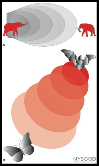
Figure 2: (A) Elephants can generate and detect the sound of frequencies In 1989, P. Ting and V. Sivagnanaratnam reported the use of B-mode ultrasonography to demonstrate the anatomy of the axilla and to observe the spread of local anesthetics during axillary brachial plexus block. In 1994, Stephan Kapral and colleagues systematically explored brachial plexus with B-mode ultrasound. Since that time, multiple teams worldwide have worked tirelessly on defining and improving the application of ultrasound imaging in regional anesthesia. Ultrasound-guided nerve blockade is currently used routinely in the practice of regional anesthesia in many centers worldwide. Here is a summary of ultrasound quick facts:
Definition of Ultrasound Sound travels as a mechanical longitudinal wave in which back-and-forth particle motion is parallel to the direction of wave travel. Ultrasound is high-frequency sound and refers to mechanical vibrations above 20 kHz. Human ears can hear sounds with frequencies between 20 Hz and 20 kHz. Elephants can generate and detect the sound with frequencies Piezoelectric Effect The piezoelectric effect is a phenomenon exhibited by the generation of an electric charge in response to a mechanical force (squeeze or stretch) applied on certain materials. Conversely, mechanical deformation can be produced when an electric field is applied to such material, also known the piezoelectric effect (Figure 3). Both natural and human-made materials, including quartz crystals and ceramic materials, can demonstrate piezoelectric properties. Recently, lead zirconate titanate has been used as piezoelectric material for medical imaging. Lead-free piezoelectric materials are also under development. Individual piezoelectric materials produce a small amount of energy. However, by stacking piezoelectric elements into layers in a transducer, the transducer can convert electric energy into mechanical oscillations more efficiently. These mechanical oscillations are then converted into electric energy. Ultrasound Terminology Period is the time it takes for one cycle to occur; the period unit of measure is the microsecond (µs). Wavelength is the length of space over which one cycle occurs; it is equal to the travel distance from the beginning to the end of one cycle. 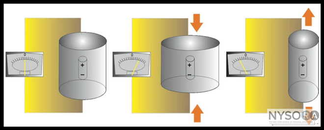
Figure 3: The piezoelectric effect. Mechanical deformation and consequent oscillation caused by an electrical field applied to certain material can produce a sound of high frequency. Acoustic velocity is the speed at which a sound wave travels through a medium. It is equal to the frequency times the wavelength. Speed is determined by the density (ρ) and stiffness (κ) of the medium (c = (κ/ρ)1/2). Density is the concentration of medium. Stiffness is the resistance of a material to compression. Propagation speed increases if the stiffness is increased or the density is decreased. The average propagation speed in soft tissues is 1540 m/s (ranges from 1400 m/s to 1640 m/s). However, ultrasound cannot penetrate lung or bone tissues. Acoustic impedance(z) is the degree of difficulty demonstrated by a sound wave being transmitted through a medium; it is equal to density ρ multiplied by acoustic velocity c (z = ρc). It increases if the propagation speed or the density of the medium is increased. Attenuation coefficient is the parameter used to estimate the decrement of ultrasound amplitude in a certain medium as a function of ultrasound frequency. The attenuation coefficient increases with increasing frequency; therefore, a practical consequence of attenuation is that the penetration decreases as frequency increases (Figure 4). Ultrasound waves have a self-focusing effect, which refers to the natural narrowing of the ultrasound beam at a certain travel distance in the ultrasonic field. It is a transition level between near field and far field. The beam width at the transi- tion level is equal to half the diameter of the transducer. At the distance of two times the near-field length, the beam width reaches the transducer diameter. The self-focusing effect amplifies ultrasound signals by increasing acoustic pressure. In ultrasound imaging, there are two aspects of spatial resolution: axial and lateral. Axial resolution is the minimum separation of above-below planes along the beam axis. It is determined by spatial pulse length, which is equal to the product of wavelength and the number of cycles within a pulse. It can be presented in the following formula: Axial resolution = wavelength (λ) × number of cycle per pulse (n) ÷ 2 The number of cycles within a pulse is determined by the damping characteristics of the transducer. The number of cycles within a pulse is usually set between 2 and 4 by the manufacturer of the ultrasound machines. As an example, if a 2-MHz ultrasound transducer is theoretically used to do the scanning, the axial resolution would be between 0.8 and 1.6 mm, making it impossible to visualize a 21-G needle. For a constant acoustic velocity, higher frequency ultrasound can detect smaller objects and provide a better resolution image. Figure 5 shows the images at different resolutions when a 0.5-mm-diameter object is visualized with three different frequency settings. Lateral resolution is another parameter of sharpness to describe the minimum side-by-side distance between two objects. It is determined by both ultrasound frequency and beam width. Lateral resolution can be improved by focusing to reduce the beam width. 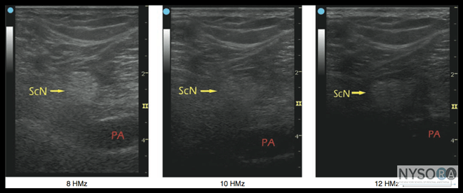
Figure 4: The ultrasound amplitude decreases in certain media as a function of ultrasound frequency, a phenomenon known as attenuation coefficient. ScN-Sciatic nerve, PA - Popliteal artery. 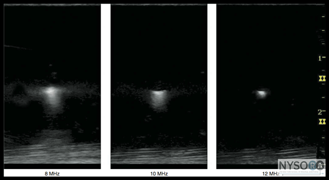
Figure 5: Ultrasound frequency affects the resolution of the imaged object. Resolution can be improved by increasing frequency and reducing the beam width by focusing. Temporal resolution is also important to observe moving objects, such as blood vessels and the heart. Similar to a movie or cartoon video, the human eye requires that the image be updated at a rate of approximately 25 times a second or higher for an ultrasound image to appear continuous. However, imaging resolution is compromised by increasing the frame rate. Optimizing the ratio of resolution to frame rate is essential to provide the best possible image. Interactions of Ultrasound Waves with Tissue 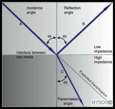
Figure 6: The interaction of ultrasound waves through the media in which they travel is complex. When ultrasound encounters boundaries between different media, part of the ultrasound is reflected and part transmitted. The reflected and transmitted directions depend on the respective angles of reflection and transmission. As the ultrasound wave travels through tissue, it is subject to a number of interactions. The most important features are as follows:
When an ultrasound wave encounters boundaries between different media, part of the ultrasound is reflected and other part is transmitted. The reflected and transmitted directions are given by the reflection angle θr and transmission angle θt, respectively (Figure 6). Reflection of a sound wave is very similar to optical reflection. Some of its energy is sent back into the originating medium. In a true reflection, reflection angle θr must equal incidence angle θi. The strength of the reflection from an interface is variable and depends on the difference of impedances between two affinitive media and the incident angle at boundary. If the media impedances are equal, there is no reflection (no echo). If there is a significant difference between media impedances, there will be far greater or nearly complete reflection. For example, an interface between soft tissues and either lung or bone involves a considerable change in acoustic impedance and creates strong echoes. This reflection intensity is also highly angle dependent, meaning that the ultrasound transducer must be placed perpendicularly to the target nerve to visualize it clearly. 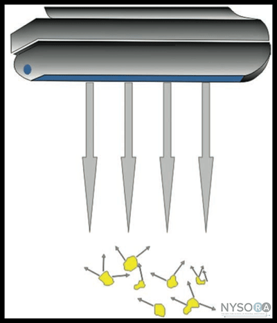
Figure 7: Scattering is the redirection of ultrasound in any direction caused by rough surfaces or by heterogeneous media. A change in sound direction when crossing the boundary between two media is called refraction. If the propagation speed through the second medium is slower than it is through the first medium, the refraction angle is smaller than the incident angle. Refraction can cause artifacts such as those that occur beneath large vessels. During ultrasound scanning, a coupling medium must be used between the transducer and the skin to displace air from the transducer-skin interface. A variety of gels and oils are applied for this purpose. They also act as lubricants, provid- ing a smooth surface scanning. Most scanned interfaces are somewhat irregular and curved. If boundary dimensions are significantly less than the wavelength or not smooth, the reflected waves will be diffused. Scattering is the redirection of sound in any direction by rough surfaces or by heterogeneous media (Figure 7). Normally, scattering intensity is much less than mirrorlike reflection intensities and is relatively independent of the direction of the incident sound wave. Therefore, the visualization of the target nerve is not significantly influenced by other nearby scattering. Absorption is defined as the direct conversion of sound energy into heat. In other words, ultrasound scanning generates heat in the tissue. Higher frequencies are absorbed at a greater rate than lower frequencies. However, higher scanning frequency gives better axial resolution. If the ultrasound penetration is not sufficient to visualize the structures of interest, a lower frequency is selected to increase the penetration. The use of longer wavelengths (lower frequency) results in lower resolution because the resolution of ultrasound imaging is proportional to the wavelength of the imaging wave. Frequencies between 6 and 12 MHz typically yield better resolution for imaging of the peripheral nerves because they are located more superficially. Lower imaging frequencies, between 2 and 5 MHz, are usually needed for imaging of neuraxial structures. For most clinical applications, frequencies less than 2 MHz or higher than 15 MHz are rarely used because of insufficient resolution or insufficient penetration, respectfully. Ultrasound Image Modes A-Mode The A-mode is the oldest ultrasound modality, dating back to 1930. The transducer sends a single pulse of ultrasound into the medium and waits for the returned signal. Consequently, a simple one-dimensional ultrasound image is generated as a series of vertical peaks corresponding to the depth of the structures at which the ultrasound beam encounters different tissues. The distance between the echoed spikes (Figure 8) can be calculated by dividing the speed of the ultrasound in the tissue (1540 m/sec) by half the elapsed time. This mode provides little information on the spatial relationships of the imaged structures, however. Therefore, A-mode ultrasound is not used in regional anesthesia. B-Mode 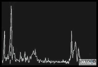
Figure 8: The A-mode of ultrasound consists of a one- dimensional ultrasound image displayed as a series of vertical peaks corresponding to the depth of structures at which the ultrasound encounters different tissues. The B-mode supplies a two-dimensional image of the area by simultaneously scanning from a linear array of 100-300 piezoelectric elements rather than a single one, as is the case in A-mode. The amplitude of the echo from a series of A-scans is converted into dots of different brightness in B-mode imaging. The horizontal and vertical directions represent real distances in tissue, whereas the intensity of the grayscale indicates echo strength (Figure 9). B mode can provide a cross sectional image through the area of interest and is the primary mode currently used in regional anesthesia. Doppler-Mode The Doppler effect is based on the work of Austrian physicist Johann Christian Doppler. The term describes a change in the frequency or wavelength of a sound wave resulting from relative motion between the sound source and the sound receiver. In other words, at a stationary position, the sound frequency is constant. If the sound source moves toward the sound receiver, a higher pitched sound occurs. If the sound source moves away from the receiver, the received sound has a lower pitch (Figure 10). Color Doppler produces a color-coded map of Doppler shifts superimposed onto a B-mode ultrasound image. Blood flow direction depends on whether the motion is toward or away from the transducer. Selected by convention, red and blue colors provide information about the direction and velocity of the blood flow. According to the color map (color bar) in the upper left-hand corner of the figure (Figure 11), the red color on the top of the bar denotes the flow coming toward the ultrasound probe, and the blue color on the bottom of the bar indicates the flow away from the probe. In ultrasound-guided peripheral nerve blocks, color Doppler mode is used to detect the presence and nature of the blood vessels (artery vs. vein) in the area of interest. When the direction of the ultrasound beam changes, the color of the arterial flow switches from blue to red, or vice versa, depending on the convention used. Power Doppler is up to five times more sensitive in detecting blood flow than color Doppler and it is less dependent on the scanning angle. Thus power Doppler can be used to identify the smaller blood vessels more reliably. The drawback is that power Doppler does not provide any information on the direction and speed of blood flow (Figure 12). 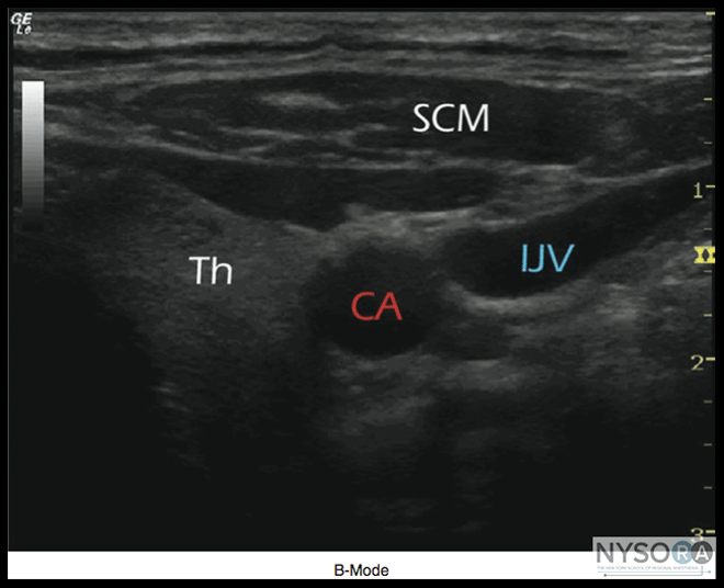
Figure 9: An example of B-mode imaging. The horizontal and vertical directions rep- resent distances and tissues, whereas the intensity of the grayscale indicates echo strength. Scm-sternocleidomastoid muscle, IJV-Internal jugular vein, CA-carotid artery, Th-Thyroid gland. M-Mode 
Figure 10: The Doppler effect. When a sound source moves away from the receiver, the received sound has a lower pitch and vice versa. A single beam in an ultrasound scan can be used to produce a picture with a motion signal, where movement of a structure such as a heart valve can be depicted in a wave-like manner. M-mode is used extensively in cardiac and fetal cardiac imaging; however, its present use in regional anesthesia is negligible (Figure 13). Ultrasound Instruments Ultrasound machines convert the echoes received by the transducer into visible dots, which form the anatomic image on ultrasound screen. The brightness of each dot corresponds to the echo strength, producing what is known as a grayscale image. Two types of scan transducers are used in regional anesthesia: linear and curved. A linear transducer can produce parallel scan lines and rectangular display, called a linear scan, whereas a curved transducer yields a curvilinear scan and arc-shaped image (Figure 14A and B). In clinical scanning, even a very thin layer of air between the transducer and skin may reflect virtually all the ultrasound, hindering any penetration into the tissue. Therefore, a coupling medium, usually an aqueous gel, is applied between surfaces of the transducer and skin to eliminate the air layer. The ultrasound machines currently used in regional anesthesia provide a two-dimensional image, or "slice." Machines capable of producing three-dimensional images have recently been developed. Theoretically, three-dimensional (3D) imaging should help in understanding the relationship of anatomic structures and spread of local anesthetics. (Figure 14C and D). At present, however, 3D-real time imaging systems still lack the resolution and simplicity of 2D images, so their practical use in regional anesthesia is limited. 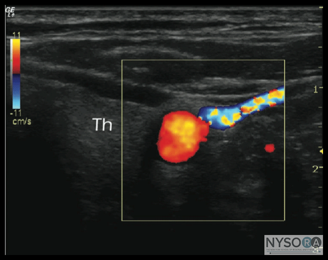
Figure 11: Color Doppler produces a color-coded map of Doppler shapes super- imposed onto a B-mode ultrasound image. Selected by convention, red and blue colors provide information about the direction and velocity of the blood flow. 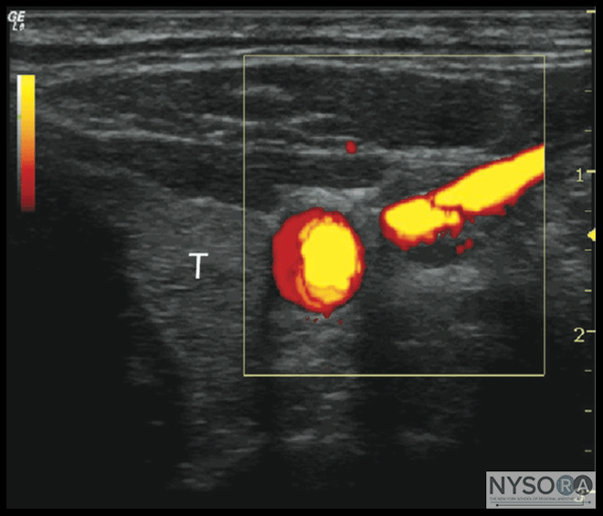
Figure 12: Although the power Doppler may be useful in identifying smaller blood vessels, the drawback is that it does not provided information on the direction and speed of blood flow. 
Figure 13: M-mode consists of a single beam used to produce an image with a motion signal. Movement of a structure can be depicted in a wavelike matter. 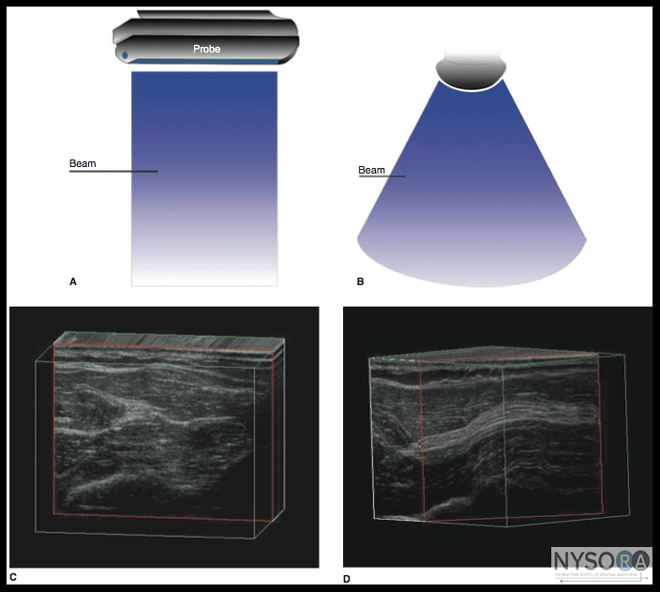
Figure 14: Three-dimensional imaging. (A) A linear transducer produces parallel scan lines and rectangular display; linear scan. (B) A curve "phase array" transducer results in a curvilinear scan and an arch-shaped image. (C) An example of cross-sectional three-dimensional (3D) imaging. (D) An example of longitudinal 3D imaging. Three-dimensional imaging theoretically should provide more spatial orientation of the image structures; however, its current drawback is lower resolution and greater complexity compared with 2D images, which limit its application in the current practice of regional anesthesia. Time-Gain Compensation The echoes exhibit a steady decline in amplitude with increasing depth. This occurs for two reasons: First, each successive reflection removes a certain amount of energy from the pulse, decreasing the generation of later echoes. Second, tissue absorbs ultrasound, so there is a steady loss of energy as the ultrasound pulse travels through the tissues. This can be corrected by manipulating time-gain compensation (TGC) and compression functions. Amplification is the conversion of the small voltages received from the transducer into larger ones that are suitable for further processing and storage. Gain is the ratio of output to input electric power. TGC is time-dependent exponential amplification. TGC function can be used to increase the amplitude of incoming signals from various tissue depths. The layout of the TGC controls varies from one machine to another. A popular design is a set of slider knobs; each knob in the slider set controls the gain for a specific depth, which allows for a well-balanced gain scale on the image (Figure 15A-C). Compression is the process of decreasing the differences between the smallest and largest echo-voltage amplitudes; the optimal compression is between 2 and 4 for maximal scale equal to 6. 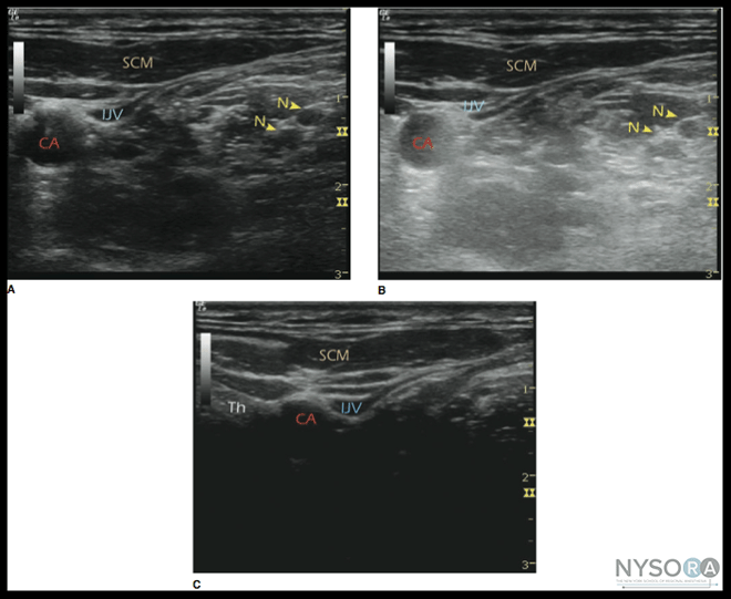
Figure 15: (A-C) The effect of the time-gain compensation settings. Time-gain compensation is a function that allows time (depth) dependent amplification of signals returning from different depths. SCM-sternocleidomastoid muscle, IJV-Internal jugular vein, N-nerve, CA-carotid artery, Th-thyroid gland. Focusing 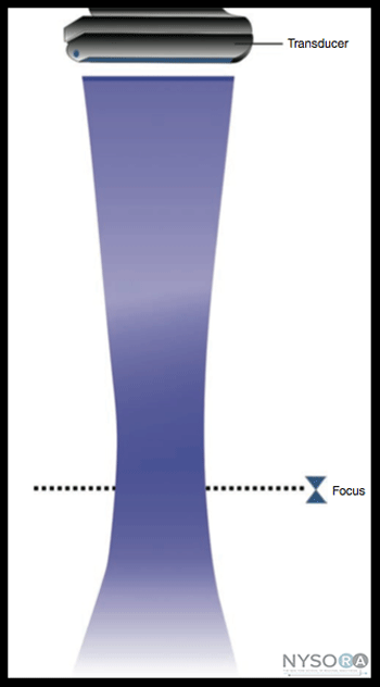
Figure 16: A demonstration of focusing effect. An electronic means can be used to narrow the width of the beam at specific depth resulting in the focusing effect and a greater resolution at a chosen depth. As previously discussed, it is common to use an electronic means to narrow the width of the beam at some depth and achieve a focusing effect similar to that obtained using a convex lens (Figure 16). This strategy improves the resolution in the plane because the beam width is converged. However, the reduction in beam width at the selected depth is achieved at the expense of degradation in beam width at other depths, resulting in poorer images below the focal region. Bioeffect and Safety The mechanisms of action by which ultrasound application could produce a biologic effect can be characterized into two aspects: heating and mechanical. The generation of heat increases as ultrasound intensity or frequency is increased. For similar exposure conditions, the expected temperature increase in bone is significantly greater than in soft tissues. Reports in animal models (mice and rats) suggest that application of ultrasound may result in a number of undesired effects, such as fetal weight reduction, postpartum mortality, fetal abnormalities, tissue lesions, hind limb paralysis, blood flow stasis, and tumor regression. Other reported undesired effects in mice are abnormalities in B-cell development and ovulatory response, and teratogenicity. In general, adult tissues are more tolerant of rising temperature than fetal and neonatal tissues. A modern ultrasound machine displays two standard indices: thermal and mechanical. The thermal index (TI) is defined as the transducer acoustic output power divided by the estimated power required to raise tissue temperature by 1°C. Mechanical index (MI) is equal to the peak rarefactional pressure divided by the square root of the center frequency of the pulse bandwidth. TI and MI indicate the relative likelihood of thermal and mechanical hazard in vivo, respectively. Either TI or MI >1.0 is hazardous. Biologic effect due to ultrasound also depends on tissue exposure time. Fortunately, ultrasound-guided nerve block requires the use of only low TI and MI values on the patient for a short period of time. Based on in vitro and in vivo experimental study results to date, there is no evidence that the use of diagnostic ultrasound in routine clinical practice is associated with any biologic risks.
|
| 02/20/2016(+ 2016 Dates) | |
| 01/27/2016 | |
| 03/17/2016 | |
| 03/23/2016 (+ 2016 Dates) | |
| 04/20/2016 | |
| 09/23/2016 | |
| 10/01/2024 |
![[advertisement] gehealthcare](../../files/banners/banner1_250x600/GEtouch(250X600).gif)




Post your comment