Essentials of Regional Anesthesia Anatomy
--- Authors: Admir Hadzic and Carlo Franco
|
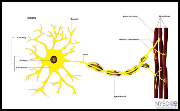
A good practical knowledge of anatomy is important for the successful and safe practice of regional anesthesia. In fact, just as surgical disciplines rely on surgical anatomy, regional anesthesiologists need to have a working knowl- edge of the anatomy of nerves and associated structures that does not include unnecessary details. In this chapter, the basics of regional anesthesia anatomy necessary for successful implementation of various techniques described later in the book are outlined. Anatomy of Peripheral Nerves All peripheral nerves are similar in structure. The neuron is the basic functional unit responsible for the conduction of nerve impulses (Figure 1). Neurons are the longest cells in the body, many reaching a meter in length. Most neurons areincapable of dividing under normal circumstances, and they have a very limited ability to repair themselves after injury. A typical neuron consists of a cell body (soma) that contains a large nucleus. The cell body is attached to several branching processes, called dendrites, and a single axon. Dendrites receive incoming messages; axons conduct outgoing messages. Axons vary in length, and there is only one per neuron. In peripheral nerves, axons are very long and slender. They are also called nerve fibers. Connective Tissue
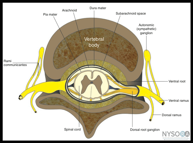
Figure 1: Organization of the peripheral nerve 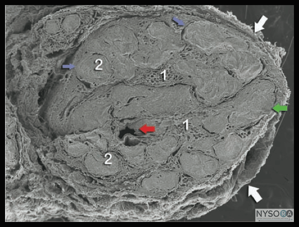 Figure 2: Histology of the peripheral nerve and connective tissues. White arrows: External epineurium (epineural sheath), 1 = Internal epineurium, 2 = fascicles, Blue arrows: Perineurium, Red arrow: Nerve vasculature Green arrow: Fascicular bundle. The individual nerve fibers that make up a nerve, like individual wires in an electric cable, are bundled together by connective tissue. The connective tissue of a peripheral nerve is an important part of the nerve. According to its position in the nerve architecture, the connective tissue is called the epineurium, perineurium, or endoneurium (Figure 2). The epineurium surrounds the entire nerve and holds it loosely to the connective tissue through which it runs. Each group of axons that bundles together within a nerve forms a fascicle, which is surrounded by perineurium. It is at this level that the nerve–blood barrier is located and constitutes the last protective barrier of the nerve tissue. The endoneurium is the fine connective tissue within a fascicle that surrounds every individual nerve fiber or axon. Nerves receive blood from the adjacent blood vessels running along their course. These feeding branches to larger nerves are macroscopic and irregularly arranged, forming anastomoses to become longitudinally running vessel(s) that supply the nerve and give off subsidiary branches. Organization of the Spinal Nerves The nervous system consists of central and peripheral parts. The central nervous system includes the brain and spinal cord. The peripheral nervous system consists of the spinal, cranial, and autonomic nerves, and their associated ganglia. Nerves are bundles of nerve fibers that lie outside the central nervous system and serve to conduct electrical impulses from one region of the body to another. The nerves that make their exit through the skull are known as cranial nerves, and there are 12 pairs of them. The nerves that exit below the skull and between the vertebrae are called spinal nerves, and there are 31 pairs of them. Every spinal nerve has its regional number and can be identified by its association with the adjacent vertebrae (Figure 3). In the cervical region, the first pair of spinal nerves, C1, exits between the skull and the first cervical vertebra. For this reason, a cervical spinal nerve takes its name from the vertebra below it. In other words, cervical nerve C2 precedes vertebra C2, and the same system is used for the rest of the cervical series. The transition from this identification method occurs between the last cervical and first thoracic vertebra. The spinal nerve lying between these two vertebrae has been designated C8. Thus there are seven cervical vertebrae but eight cervical nerves. Spinal nerves caudal to the first thoracic vertebra take their names from the vertebra immediately preceding them. For instance, the spinal nerve T1 emerges immediately caudal to vertebra T1, spinal nerve T2 passes under vertebra T2 and so on.
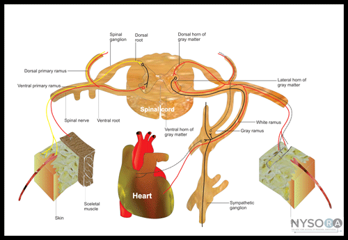
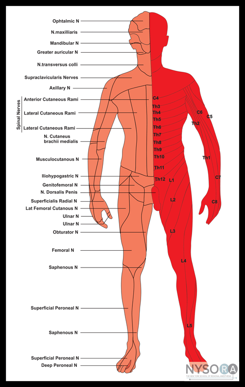
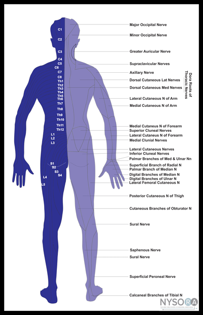
Figure 3: Organization of the spinal nerve Connective Tissue Each spinal nerve is formed by a dorsal and a ventral root that come together at the level of the intervertebral fora- men (Figure 3). In the thoracic and lumbar levels, the first branch of the spinal nerve carries visceral motor fibers to a nearby autonomic ganglion. Because preganglionic fibers are myelinated, they have a light color and are known as white rami (Figure 4). Two groups of unmyelinated postganglionic fibers leave the ganglion. Those fibers innervating glands and smooth muscle in the body wall or limbs form the gray ramus that rejoins the spinal nerve. The gray and white rami are collectively called the rami communi- cantes. Preganglionic or postganglionic fibers that inner- vate internal organs do not rejoin the spinal nerves. Instead, they form a series of separate autonomic nerves and serve to regulate the activities of organs in the abdominal and pelvic cavities. The dorsal ramus of each spinal nerve carries sensory innervation from, and motor innervation to, a specific segment of the skin and muscles of the back. The region innervated resembles a horizontal band that begins at the origin of the spinal nerve. The relatively larger ventral ramus supplies the ventrolateral body surface, structures in the body wall, and the limbs. Each spinal nerve supplies a specific segment of the body surface, known as a dermatome. Dermatomes A dermatome is an area of the skin supplied by the dorsal (sensory) root of the spinal nerve (Figures 5 and 6). In the head and trunk, each segment is horizontally disposed, except C1, which does not have a sensory component.
Figure 4: Organization and function of the segmental (spinal nerve). The dermatomes of the limbs from the fifth cervical to the first thoracic nerve, and from the third lumbar to the second sacral vertebrae, form a more complicated arrangement due to rotation and growth during embryologic life. There is considerable overlapping of adjacent dermatomes; that is, each segmental nerve overlaps the territories of its neighbors. This pattern is variable among individuals, and it is more of a guide than a fixed map.
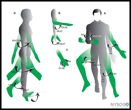
Myotomes A myotome is the segmental innervation of skeletal muscle by a ventral root of a specific spinal nerve (Figure 7).
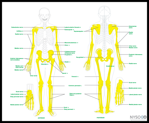
Figure 7: Motor innervation of the major muscle groups. (A) Medial and lateral rotation of shoulder and hip. Abduction and adduction of shoulder and hip. (B) Flexion and extension of elbow and wrist. (C) Pronation and supination of forearm. (D) Flexion and extension of shoulder, hip, and knee. Dorsiflexion and plantar flexion of ankle, lateral views. Osteotomes The innervation of the bones follows its own pattern and does not coincide with the innervation of more superficial structures (Figure 8).
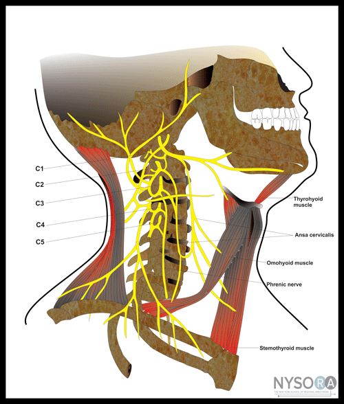
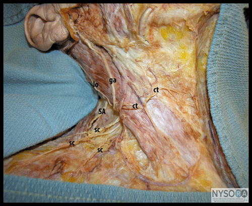
Figure 8: Osteotomes Nerve Plexuses Although the dermatomal innervation of the trunk is simple, the innervation of the extremities, part of the neck, and pelvis is highly complex. In these areas, the ventral rami of the spinal nerves form an intricate neural network; nerve fibers coming from similar spinal segments easily reach different terminal nerves. The four major nerve plexuses are the cervical plexus, brachial plexus, lumbar plexus, and sacral plexus. The Cervical Plexus The cervical plexus originates from the ventral rami of C1-C5, which form three loops (Figures 9 and 10). Branches from the cervical plexus provide sensory innervation of part of the scalp, neck, and upper shoulder and motor innervation to some of the muscles of the neck, the thoracic cavity, and the skin (Table 1). The phrenic nerve, one of the larger branches of the plexus, innervates the diaphragm.
Figure 9: Organization of the cervical plexus
Figure 10: Superficial cervical plexus branches. ct, transverse cervical; ga, greater auricular; lo, lesser occipital; sc, supra- clavicular. Also shown is the spinal accessory nerve (SA). Table 1: Organization and Distribution of the Cervical Plexus
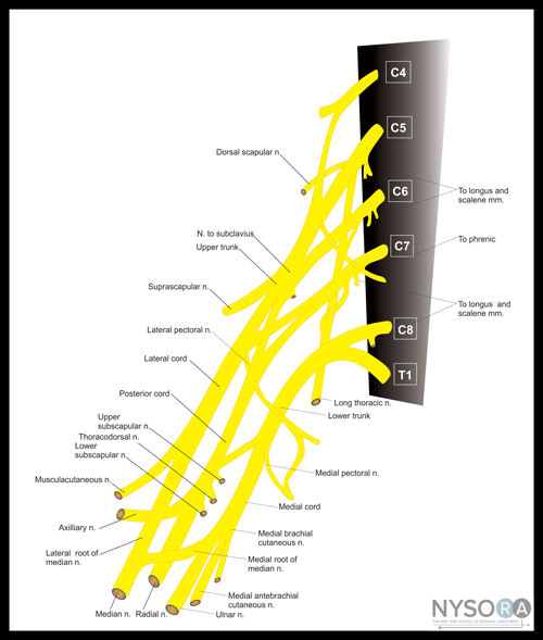
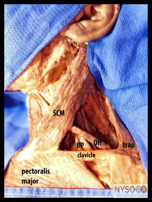
The Brachial Plexus The brachial plexus is both larger and more complex than the cervical plexus (Figures 11, 12, 13, 14A,B, 15A,B, and 16). It innervates the pectoral girdle and upper limb. The plexus is formed by five roots that originate from the ventral rami of spinal nerves C5-T1. The roots converge to form the superior (C5-C6), middle (C7), and inferior (C8-T1) trunks (Table 2). The trunks give off three anterior and three posterior divisions as they approach the clavicle. The divisions rearrange their fibers to form the lateral, medial, and posterior cords. The cords give off the terminal branches. The lateral cord gives off the musculocutaneous nerve, and the lateral root of the median nerve. The medial cord gives off the medial root of the median nerve and the ulnar nerve. The posterior cord gives off the axillary and radial nerves.
Figure 11: Organizatio of the brachial plexus
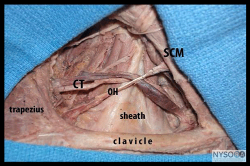
Figure 14: (A) A thick fascia layer (sheath) covers the brachial plexus in the posterior triangle. Also seen is part of the sternocleidomastoid muscle (SCM), the cervical transverse vessels (CT), and the omohyoid muscle (OH). (B) Once the sheath is removed, the brachial plexus can be seen between the anterior scalene (AS) and middle scalene (MS) muscles.
Figure 16: Intercostobrachial nerve (T2) is the lateral branch of the second intercostal nerve that supplies sensory innerva- tion to the axilla and upper medial side of the arm. Table 2: Organization and Distribution of the Brachial Plexus
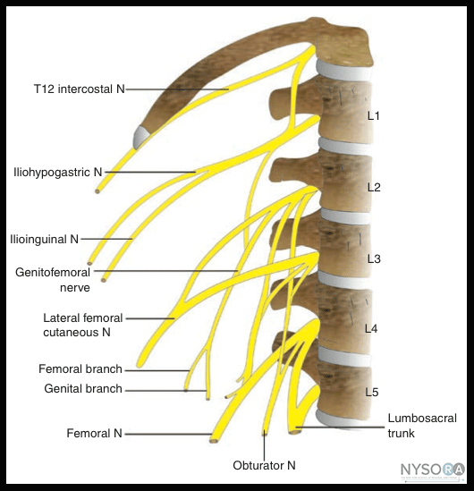
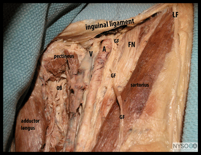
The Lumbar Plexus The lumbar plexus is formed by the ventral rami of spinal nerves L1-L3 and the superior branch of L4 (Figures 17, 18A,B, and 19). In about 50% of the cases, there is a contribution from T12. The inferior branch of L4, along with the entire ventral rami of L5, forms the lumbosacral trunk that contributes to the sacral plexus. Because the branches of both the lumbar and sacral plexuses are distributed to the lower limb, they are often collectively referred to as the lumbosacral plexus. The main branches of the lumbar plexus are the iliohypogastric, ilioinguinal, genitofemoral, lateral femoral cutaneous, obturator, and femoral nerves (Figures 19, 20A,B; Table 3).
Figure 17: Intercostobrachial nerve (T2) is the lateral branch of the second intercostal nerve that supplies sensory innerva- tion to the axilla and upper medial side of the arm.
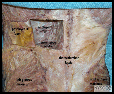
Figure 18: (A) Posterior view of the back to show the thoracolumbar fascia (TLF), whose posterior layer has been open as a small window through which part of the erector spinae muscles has been resected to show the anterior layer of the thoracolumbar fascia. (B) One step further in the dissection shows part of the quadratus lumborum muscle.
Figure 19: Two branches of the lumbar plexus, the femoral nerve and obturator, are seen between the quadratus lumborum and psoas muscles in the right retroperitoneal space. Table 3: Organization and Distribution of the Lumbar Plexus
Figure 20:(A) Frontal view of the upper anterior thigh showing the inguinal ligament and some branches of the lumbar plexus: FN, femoral nerve; GF, femoral branch of genitofemoral nerve; LF, lateral femoral cutaneous nerve; OB, obturator nerve. The femoral vein (V) and artery (A) are also shown. (B) The same nerves of (A) are shown from the lateral side.
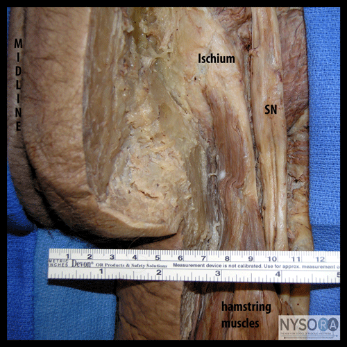
The Sacral Plexus The sacral plexus arises from the lumbosacral trunk (L4-L5) plus the ventral rami of S1-S4 (Figures 21, 22A,B, 23, and 24). The main nerves of the sacral plexus are the sciatic nerve and the pudendal nerve (Table 4). The sciatic nerve leaves the pelvis through the greater sciatic foramen to enter the gluteal area where it travels between the greater trochanter and ischial tuberosity. In the proximal thigh it lies behind the lesser trochanter of the femur covered superficially by the long head of the biceps femoris muscle. As it approaches the popliteal fossa, the two components of the sciatic nerve diverge into two recognizable nerves: the common peroneal and the tibial nerve (Figures 25 and 26).
Figure 21: Organization of the sacral plexus
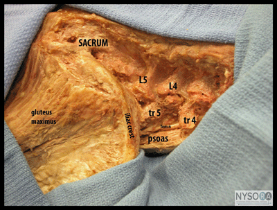
Figure 22: (A) The back and paraspinal muscles have been removed to show the transverse processes of the last lumbar vertebra, the psoas muscle, and the femoral nerve. (B) Same as (A) showing that the lateral edge of the psoas muscle at the iliac crest is between 4 and 5 cm from the midline. fem, femoral.
Table 4: Organization and Distribution of the Sacral Plexus
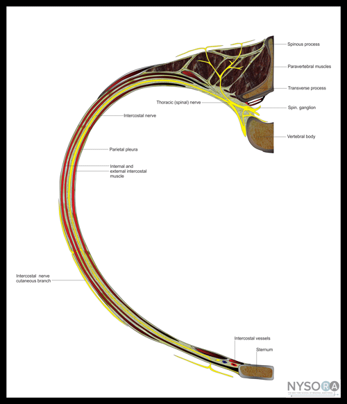
Thoracic and Abdominal Wall Thoracic Wall The intercostal nerves originate from the ventral rami of the first 11 thoracic spinal nerves. Each intercostal nerve becomes part of the neurovascular bundle of the rib and provides sensory and motor innervations (Figure 27). Except for the first, each intercostal nerve gives off a lateral cutaneous branch that pierces the overlying muscle near the midaxillary line. This cutaneous nerve divides into anterior and posterior branches, which supply the adjacent skin. The intercostal nerves of the second to the sixth spaces reach the anterior thoracic wall and pierce the superficial fascia near the lateral border of the sternum and divide into medial and lateral cutaneous branches. Most of the fibers of the anterior ramus of the first thoracic spinal nerve join the brachial plexus for distribution to the upper limb. The small first intercostal nerve is in itself the lateral branch and supplies only the muscles of the intercostal space, not the overlying skin. The lower five intercostal nerves abandon the intercostal space at the costal margin to supply the muscles and skin of the abdominal wall. Anterior Abdominal Wall The skin, muscles and parietal peritoneum, or the anterior abdominal wall, are innervated by the lower six thoracic nerves and the first lumbar nerve. At the costal margin, the seventh to eleventh thoracic nerves leave their intercostal spaces and enter the abdominal wall in a fascial plane between the transversus abdominis and internal oblique muscles. The seventh and eighth intercostal nerves slope upward following the contour of the costal margin, the ninth runs horizontally, and the tenth and eleventh have a somewhat downward trajectory. Anteriorly, the nerves pierce the rectus abdominis muscle and the anterior layer of the rectus sheath to emerge as anterior cutaneous branches that supply the overlying skin. The subcostal nerve (T12) takes the line of the twelfth rib across the posterior abdominal wall. It continues around the flank and terminates in a similar manner to the lower intercostal nerves. The seventh to twelfth thoracic nerves give off lateral cutaneous nerves that further divide into anterior and posterior branches. The anterior branches supply the skin as far forward as the lateral edge of rectus abdominis. The posterior branches supply the skin overlying the latissimus dorsi. The lateral cutaneous branch of the subcostal nerve is distributed to the skin on the side of the buttock.
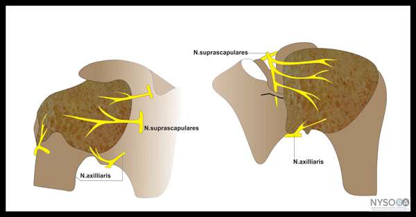
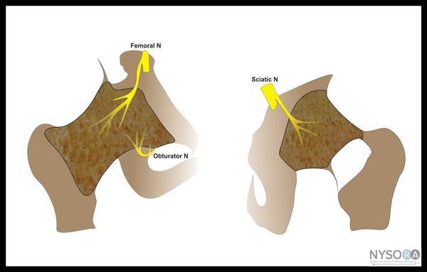
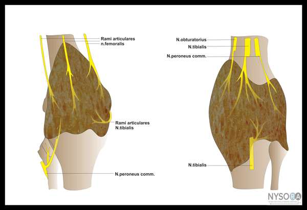
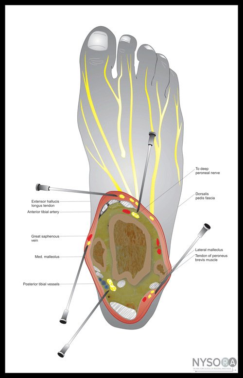
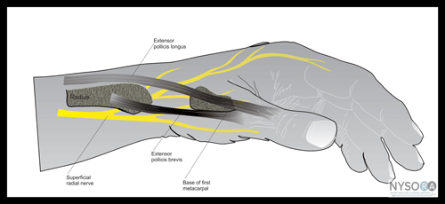
Figure 27: Organization of the segmental spinal nerve, intercostal nerve, and innervations of the chest wall. The inferior part of the abdominal wall is supplied by the iliohypogastric and ilioinguinal nerves, both branches of L1.The iliohypogastric nerve divides, runs above the iliac crest, and splits into two terminal branches. The lateral cutaneous branch supplies the side of the buttock; the anterior cutaneous branch supplies the suprapubic region. The ilioinguinal nerve leaves the intermuscular plane by piercing the internal oblique muscle above the iliac crest. It continues between the two oblique muscles eventually to enter the inguinal canal through the spermatic cord. Emerging from the superficial inguinal ring, it gives cutaneous branches to the skin on the medial side of the root of the thigh, the proximal part of the penis, and the front of the scrotum in males and the mons pubis and the anterior part of the labium majus in females. Nerve Supply to the Peritoneum The parietal peritoneum of the abdominal wall is innervated by the lower thoracic and first lumbar nerves. The lower thoracic nerves also innervate the peritoneum that covers the periphery of the diaphragm. Inflammation of the peritoneum gives rise to pain in the lower thoracic wall and abdominal wall. By contrast, the peritoneum on the central part of the diaphragm receives sensory branches from the phrenic nerves (C3, C4, and C5), and irritation in this area may produce pain referred to region of the shoulder (the fourth cervical dermatome). Innervation of the Major Joints Because much of the practice of peripheral nerve blocks involves orthopedic surgery, it is important to review the innervation of the major joints to have a better understanding of the nerves involved for a more rational approach to regional anesthesia. Shoulder Joint Innervation to the shoulder joints originates mostly from the axillary and suprascapular nerves, both of which can be blocked by an interscalene block (Figure-28). Elbow Joint Nerve supply to the elbow joint includes branches of all major nerves of the brachial plexus: musculocutaneous, radial, median, and ulnar nerves. Hip Joint Nerves to the hip joint include the nerve to the rectus femoris from the femoral nerve, branches from the anterior division of the obturator nerve, and the nerve to the quadratus femoris from the sacral plexus (Figure 29). Knee Joint The knee joint is innervated anteriorly by branches from the femoral nerve. On its medial side it receives branches from the posterior division of the obturator nerve while both divisions of the sciatic nerve supply its posterior side (Figure 30). Ankle Joint The innervation of the ankle joint is complex and involves the terminal branches of the common peroneal (deep and superficial peroneal nerves), tibial (posterior tibial nerve), and femoral nerves (saphenous nerve). A more simplistic view is that the entire innervation of the ankle joint stems from the sciatic nerve, with the exception of the skin on the medial aspect around the medial malleolus (saphenous nerve, a branch of the femoral nerve) (Figure 31). Wrist Joint The wrist joint and joints in the hand are innervated by most of the terminal branches of the brachial plexus including the radial, median, and ulnar nerves (Figure 32).
Figure 28: Innervation of the shoulder joint
Figure 29:Innervation of the hip joint
Figure 30: Innervation of the knee joint.
Figure 31: Innervation of the ankle joint and foot
Figure 32: Innervation of the wrist and hand
|
| 12/19/2015(+ 2016 Dates) | |
| 01/27/2016 | |
| 03/17/2016 | |
| 04/20/2016 | |
| 09/24/2016 | |
| 10/01/2024 |
![[advertisement] gehealthcare](../files/banners/banner1_250x600/GEtouch(250X600).gif)

![[advertisement] concertmedical](../files/bk-nysora-ad.jpg)

 A
A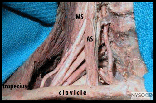 B
B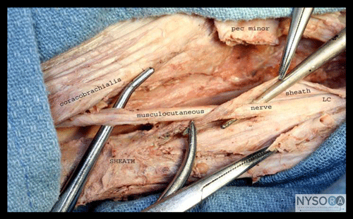 A
A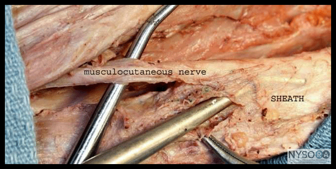 B
B
 A
A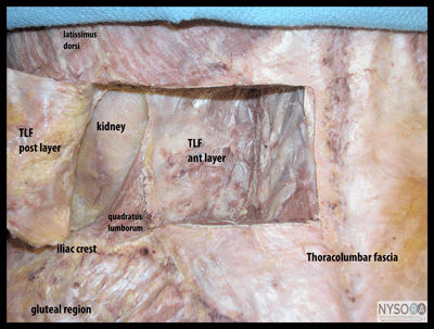 B
B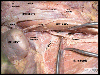
 A
A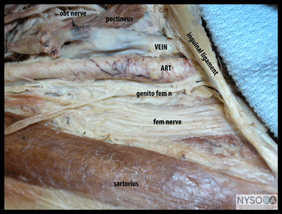 B
B
 A
A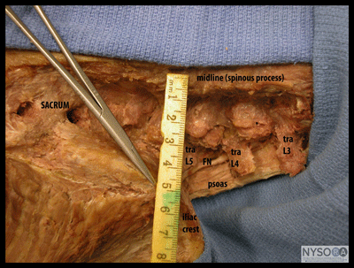 B
B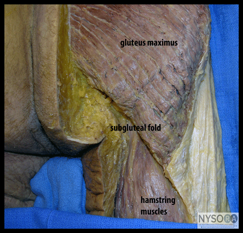





Post your comment