Lumbar Plexus Block
|
Authors: Manoj Karmakar and Catherine Vandepitte Introduction Lumbar plexus block (LPB) traditionally is performed using surface anatomic landmarks to identify the site for needle insertion and eliciting quadriceps muscle contraction in response to nerve electrolocalizati-on, as described in the nerve stimulator-guided chapter. The main challenges in accomplishing LPB relate to the depth at which the lumbar plexus is located and the size of the plexus, which requires a large volume of local anesthetic for success. Due to the deep anatomic location of the lumbar plexus, small errors in landmark estimation or angle miscalculations during needle advancement can result in needle placement away from the plexus or at unwanted locations. Therefore, monitoring of the needle path and final needle tip placement should increase the precision of the needle placement and the delivery of the local anesthetic. Although computed tomography and fluoroscopy can be used to increase the precision during LPB, these technologies are impractical in the busy operating room environment, costly, and associated with radiation exposure. It is only logical, then, that ultrasound-guided LPB is of interest because of the ever-increasing availability of portable machines and the improvement in the quality of the images obtained. (1,2) 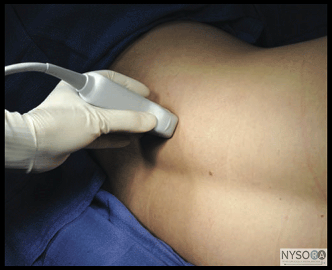
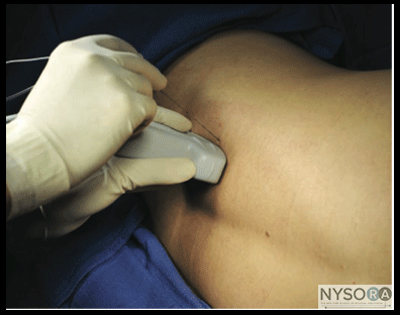
Figure 1: Transducer position (curved transducer, longitudinal view) to image the central neuroaxis, transverse processes, and estimate the needle and depth to the lumbar plexus using a longitudinal view. Anatomy and Sonoanatomy Lumbar plexus block, also known as psoas compartment block, comprises an injection of local anesthetic in the fascial plane within the posterior aspect of the psoas major muscle. Because the roots of the lumbar plexus are located in this plane, an injection of a sufficient volume of local anesthetic in the postero-medial compartment of the psoas muscle results in block of the majority of the plexus (femoral nerve, lateral femoral cutaneous nerve, and the obturator nerve). The anterior boundary of the fascial plane that contains the lumbar plexus is formed by the fascia between the anterior two thirds of the compartment of the psoas muscle that originates from the anterolateral aspect of the vertebral body and the posterior one third of the muscle that originates from the anterior aspect of the transverse processes. This arrangement explains why the transverse processes are closely related to the plexus and therefore are used as the main landmark during LPB.
A scan for the LPB can be performed in the transverse or longitudinal axes. The ultrasound transducer is positioned 3 to 4 cm lateral to the lumbar spine for either orientation. The following settings usually are used to start the scanning:
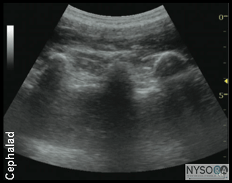
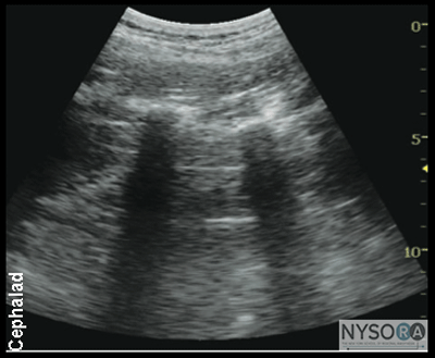
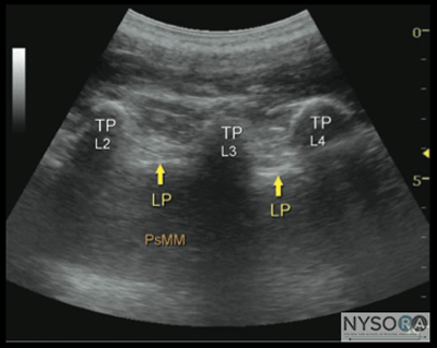
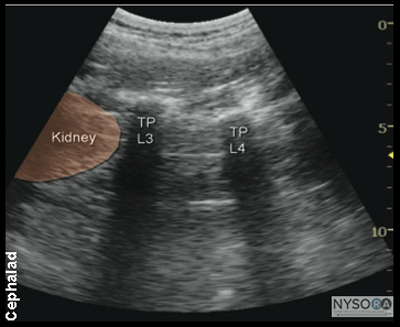
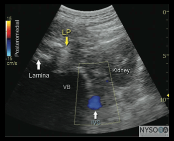
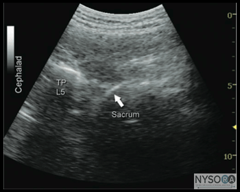
Longitudinal Scan Anatomy 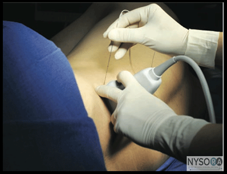 Figure 4: Patient position (lateral decubitus position) transducer (curved, linear array) placement and the needle insertion angle to block the lumbar plexus using oblique transverse view. Regardless of the technique, the operator first should identify the transverse processes on a longitudinal sonogram (Figure 1). One technique is to first identify the flat surface of the sacrum and then scan proximally until the intervertebral space between L5 and S1 is recognized as an interruption of the sacral line continuity. Once the operator identifies the transverse process of L5, the transverse process of the other lumbar vertebrae are easily identified by a dynamic cephalad scan in ascending order. The acoustic shadow of the transverse process has a characteristic appearance, often referred to as a "trident sign" (Figure 2A). Once the transverse processes are recognized, the psoas muscle is imaged through the acoustic window of the transverse processes. The psoas muscle appears as a combination of longitudinal hyperechoic striations within a typical hypoechoic muscle appearance just deep to the transverse processes (Figure 2B). Although some of the hyperechoic striations may appear particularly intense and mislead the operator to interpret them as roots of lumbar plexus, the identification of the roots in a longitudinal scan is not reliable without nerve stimulation. This unreliability is partly due to the fact that intramuscular connective tissue (e.g., septa, tendons) within the psoas muscle are thick and may be indistinguishable from the nerve roots at such a deep location. As the transducer is moved progressively cephalad, the lower pole of the kidney often comes into view as low as L2-L4 in some patients (Figure 3A and B). Transverse Scan Anatomy 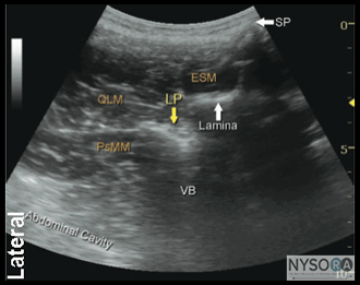 A Kirchmair and colleagues were among the first to describe the sonoanatomy of relevance for LPB. (3) They reported the ability to accurately guide a needle to the posterior part of the psoas muscle, where the roots of the lumbar plexus are located, using ultrasound guidance in cadavers. (4,5) Since, significant advances in ultrasound technology have taken place, allowing for much improved image quality, which have allowed Karmakar and colleagues to devise an alternative approach to the lumbar plexus using ultrasonographic identification of the transverse processes as the guide. (6) With this scanning technique, the transducer is positioned 4 to 5 cm lateral to the lumbar spinous process at the L3-L4 level and directed slightly medially to assume a transverse oblique orientation (Figure 4). This approach allows imaging of the lumbar paravertebral region with the erector spinae muscle, transverse process, the psoas major muscle, quadrates lumborum, and the anterolateral surface of the vertebral body (Figure 5A, B, and C). In the transverse oblique view, the inferior vena cava (IVC), on the right-sided scan, or the aorta, on the left-sided scan, also can be seen and provide additional information on the location of the psoas muscle, which is positioned superficial to these vessels. In this view, the psoas muscle appears slightly hypoechoic with multiple hyperechogenic striations within. The lower pole of the kidney can often be seen, when scanning at the L2-L4 level, as an oval structure that ascends and descends with respirations (Figure 6). The key to obtaining adequate images of the psoas muscle and lumbar plexus with the transverse oblique scan is to insonate between two adjacent transverse processes. This scanning method avoids acoustic shadow of the transverse processes, which obscures the underlying psoas muscle and the intervertebral foramen (angle between the transverse process and vertebral body) and allows visualization of the articular process of the facet joint (APFJ) as well. Because the intervertebral foramen is located at the angle between the APFJ and vertebral body, lumbar nerve roots often can be depicted.
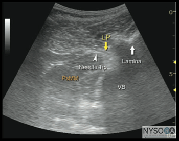
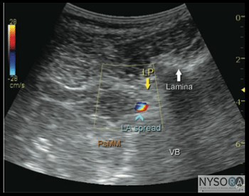
Figure 5: (A) Ultrasound anatomy of the lumbar paravertebral space using transverse oblique view. SP, spinal process; ESM, erectors spinae muscle; QLM, quadratus lumborum muscle; PsMM, psoas major muscle; VB, vertebral body. The lumbar plexus root is seen just below the lamina as it exits the interlaminar space and enters into the posterior medial aspect of the PsMM. (B) Needle path in ultrasound-guided lumbar plexus block using transverse oblique view. LP, lumbar plexus; PsMM, psoas major muscle; VB, vertebral body. (C) Spread of the local anesthetic solution with lumbar plexus block injection. Due to the deep location of the plexus, spread of the local anesthetic may not always be well seen. Color Doppler imaging can be used to help determine the location of the injectate. Techniques of Ultrasound-Guided Lumbar Plexus Block Kirchmair and colleagues suggested a paramedial sagittal scan technique with transverse scan to delineate the psoas major muscle at the L3-L5 level with the patient in the lateral position. Once a satisfactory image is obtained, the needle is inserted in-plane medial to the transducer approximately 4 cm lateral to the midline. Then the needle is advanced toward the posterior part of the lumbar plexus until the correct position is confirmed by obtaining a quadriceps motor response to nerve stimulation (1.5-2.0 mA). Needle-nerve contact and distribution of the local anesthetic is not always well seen, although lumbar plexus roots may be better visualized after the injection. Injection, dosing, and monitoring principles are the same as with the nerve stimulator-guided technique.
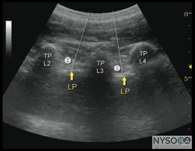
More recently, Karmakar and colleagues described the "trident sign technique," which uses an easily recognizable ultrasonographic landmark, transverse processes, and an out-of-plane needle insertion. The trident sign technique derives its name from the characteristic ultrasonographic appearance of the transverse processes (trident) to estimate the depth and location of the lumbar plexus. After application of ultrasound gel to the skin over the lumbar paravertebral region, the ultrasound transducer is positioned approximately 3 to 4 cm lateral and parallel to the lumbar spine to produce a longitudinal scan of the lumbar paravertebral region (Figure 7). Then the transducer is moved caudally, while still maintaining the same orientation, until the sacrum and the L5 transverse process become visible (Figure 8). The lumbar transverse processes are identified by their hyperechoic reflections and acoustic shadowing beneath which is typical of bone. Once the L5 transverse process is visible, the transducer is moved cephalad gradually, to identify the L3-L4 level. The goal of the technique is to guide the needle through the acoustic window between the transverse processes (between the "teeth of the trident") of L3-L4 or L2-L3 into the posterior part of the psoas major muscle containing the roots of the lumbar plexus (Figure 2B). After obtaining ipsilateral quadriceps muscle contractions, the block is carried out using the previously described injection and pharmacology considerations (Figures 9 and 10). 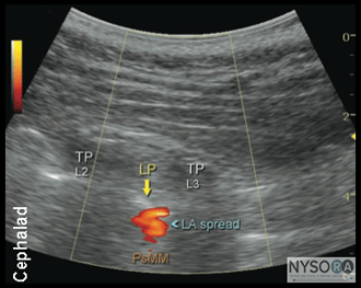 Figure 10: Local anesthetic (LA) disposition during injection of local anesthetic into the psoas muscle and the L2-L3 level. The spread of LA is often not well seen using two-dimensional imaging. LP, lumbar plexus; TP, transverse process. A paramedial scan also can be used with an in-plane needle approach. In this technique, an insulated needle is inserted in-plane from the caudal end (Figure 4) of the transducer while maintaining the view of the transverse processes. Again, the goal is to pass the needle and inject local anesthetic with a real-time visualization of the needle path and injection into the posterior part of the psoas muscle (Figure 5). In summary, ultrasound-guided LPB is a technically advanced procedure. Experience with ultrasound anatomy and less technically challenging nerve regional anesthesia techniques are useful to ensure success and safety. Although the use of ultrasound in LPB is not widely accepted, in expert hands, ultrasound guidance can increase the accuracy and possibly safety, by providing information on the location, arrangement, and depth of the osseous and muscular tissues of importance in LPB. It should be kept in mind that the dorsal branch of the lumbar artery is closely related to the trans-verse processes and the posterior part of the psoas muscle. Considering the rich vascularity of the lumbar paravertebral area, the use of smaller gauge needles and avoidance of this block in patients on anticoagulants is prudent. Injections into this area should be carried out without excessive force because high-injection pressure can lead to unwanted epidural spread and/or rapid intravascular injection. (7) Lumbar plexus block in patients with obesity or advanced age can be more challenging. Aging is associated with a reduction in skeletal muscle mass (sarcopenia) and replacement of the muscle mass by adipose tissue, leading to changes in ultrasound absorption and scattering.  |
| 12/19/2015(+ 2016 Dates) | |
| 01/27/2016 | |
| 03/17/2016 | |
| 04/20/2016 | |
| 09/24/2016 | |
| 10/01/2024 |
![[advertisement] gehealthcare](../../../files/banners/banner1_250x600/GEtouch(250X600).gif)

![[advertisement] concertmedical](../../../files/bk-nysora-ad.jpg)







Post your comment